Worm Art
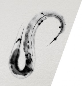
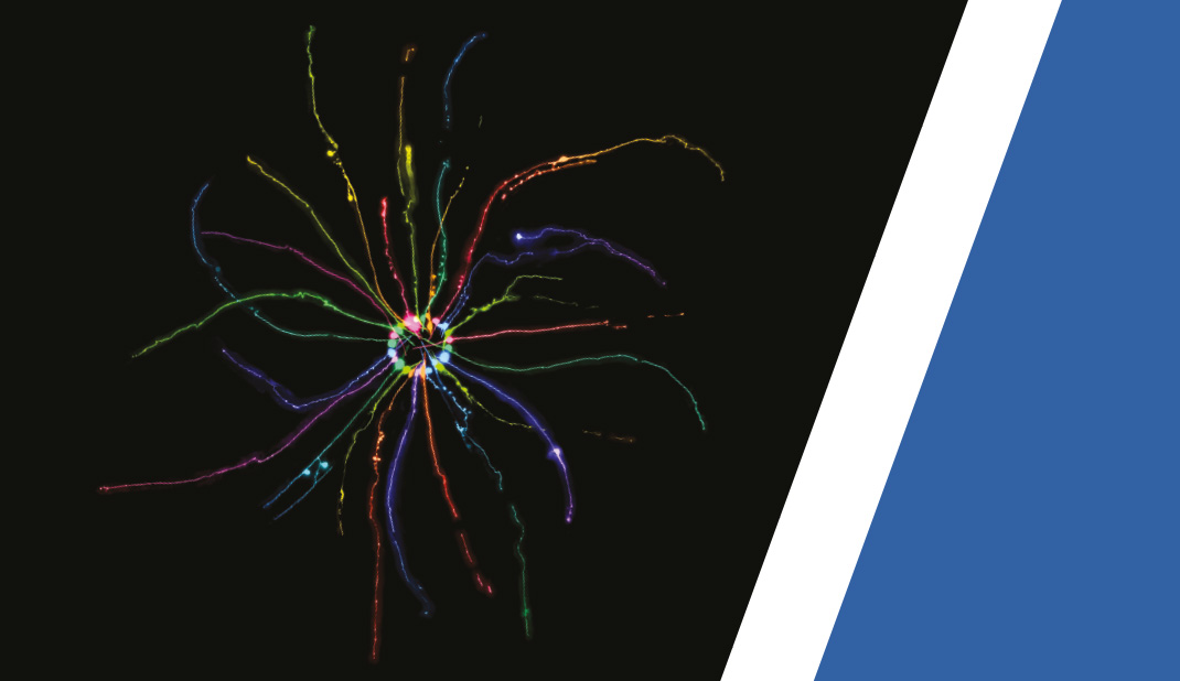
A variety of scientific images produced at the Queensland Brain Institute were used in the annual Art in Neuroscience calendars given to the staff and philanthropic donors.
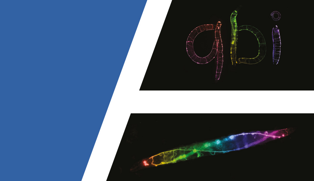
The 1.5mm-long nematodes, Caenorhabditis elegans, were imaged using fluorescence microscopy. The worms had been genetically modified so that their nerve cells fluoresced on exposure to ultraviolet light, allowing black and white micrographs to be captured.
Animals were anaesthetised prior to being imaged to minimise movement so that long exposures could be used to capture high quality images. This also allowed them to be positioned into shapes so that they could be used to form letters. Separate micrographs were digitally merged and coloured for dramatic effect.


